
Skin pigmentation changes.
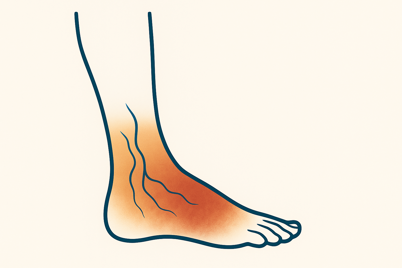
Darkening, brown patches, or blotchy skin on the legs can be an early warning sign of vein disease. Dr. Banerjee evaluates the underlying cause and treats it at the source.
- Earlysign of vein disease
- Duplexultrasound to confirm
- Treatablewhen veins are the cause
- Outpatientminimally invasive care
What is skin pigmentation?
Skin pigmentation refers to blotches or patches of discoloration on the skin. Many patients with venous insufficiency or venous stasis dermatitis notice changes in the skin around their veins. In some cases, it is the first visible warning that something deeper is off.
When the valves in your leg veins stop working properly, blood pools in the lower legs. Over time, that pooled blood breaks down and leaves behind hemosiderin, an iron rich pigment that stains the skin brown, reddish, or dark bronze.
Discoloration most often appears on the lower legs, ankles, and feet. It can show up as scattered blotches, a single dark patch, or large areas that gradually darken over months.
- Brown or reddish brown patches near the ankles
- Dark bronze coloring on the lower legs
- Shiny or thickened skin
- Itching, dryness, or tightness in the same area
- Discoloration that does not fade

More than cosmetic.
Chronic venous insufficiency, where leg vein valves stop working properly, causes blood to pool in the lower legs. Pigmentation changes often appear alongside other symptoms. If you notice any of these together, it is worth a vein evaluation.
- Enlarged or visible veins (varicose veins)
- Swelling in the legs, ankles, or feet
- Aching, heaviness, or tired legs
- Itching around discolored skin
- Restless legs, especially at night
- Cramping in the lower legs
- Non healing sores or open ulcers
- Thickened, hardened, or shiny skin
Pooled blood under the skin.
In most cases the pigment you see is hemosiderin, a byproduct of red blood cells that have leaked from overloaded veins into surrounding tissue. The skin is reacting to a circulation problem, not a skin condition.
The underlying issue is usually venous reflux. The one way valves inside your leg veins weaken, blood flows backward, and pressure builds in the lower legs. Red blood cells push out of the capillaries and break down. The iron they leave behind, hemosiderin, settles in the skin as a brown or bronze stain.
Left untreated, this often progresses to venous stasis dermatitis, where the skin becomes inflamed, itchy, and fragile. From there it can advance to open venous ulcers that are slow to heal.
Several factors raise your risk:
- Aging and family history of vein disease
- Pregnancy and hormonal changes
- Long periods of standing or sitting
- Carrying extra weight
- Prior blood clots or leg injuries
Treating the vein, not the stain.
Topical creams do not reach the underlying problem. To stop pigmentation from spreading and to let the skin recover, we treat the diseased vein that caused it. Dr. Banerjee tailors the plan after a duplex ultrasound and physical exam.
-
01
Radiofrequency Ablation
Heat closes the diseased vein. Outpatient, local anesthesia, walk in walk out.
-
02
VenaSeal
Medical adhesive seals the vein. No heat. No tumescent anesthesia. Twenty to forty minutes.
-
03
Ultrasound guided sclerotherapy
An injectable foam closes the vein from the inside. Used alone or alongside other procedures.
-
04
Lifestyle support
Compression, movement, weight management, and reduced sodium intake support healing alongside treatment.
Why choose Advanced Vascular?
- ·
Catch vein disease early. Pigmentation often shows up before pain, ulcers, or visible varicose veins.
- ·
Reduce ulcer risk. Treating the underlying reflux lowers the chance of skin breaking down into a slow healing wound.
- ·
Calm itching and inflammation. Patients often notice less itching, dryness, and irritation after treatment.
- ·
Insurance often covers it. Medically necessary vein treatment is covered by Medicare and most major insurance plans.
- ·
Skin can improve over time. With the reflux corrected, many patients see gradual fading of brown patches, though longstanding pigment may not fully clear.
- ·
Read by a vein specialist. Pigment changes point to venous pressure rather than a cosmetic issue. Review Dr. Banerjee’s credentials before your consultation.
Frequently asked questions.
If you have a question we did not cover, the team is happy to walk you through it on the phone.
Most leg pigmentation we see is caused by chronic venous insufficiency. Blood pools in the lower legs, red blood cells leak into the tissue, and the iron they release stains the skin brown, reddish, or bronze. Other causes can include skin conditions, injury, or medication, which is why an exam matters.
Treating the underlying vein often slows or stops new pigmentation and can lead to gradual fading. Longstanding or deeply set discoloration may not fully clear. The main goal is to protect the skin from worsening damage and venous ulcers. Individual results vary.
Dr. Banerjee reviews your medical history, examines the affected skin, and uses a duplex ultrasound to look at blood flow and valve function in the legs. The ultrasound is the key test for confirming venous reflux.
Sooner is better. If you notice new brown patches, darkening around the ankles, skin that itches or thickens, swelling, or any open sore on the lower leg, schedule an evaluation. Pigmentation that is ignored can progress to venous stasis dermatitis and ulcers.
Skin care
at Advanced Vascular.
Schedule an evaluation with Dr. Banerjee. A duplex ultrasound and an honest conversation about what is causing the change in your skin.